41 fluorescent labels and light microscopy
Label-free imaging of live cells | CytoSMART A non-invasive and non-toxic alternative to fluorescent microscopy is label-free imaging. Here, a combination of contrast-improving optics and image analysis algorithms are used to analyze cell cultures. ... Thorn, K. A quick guide to light microscopy in cell biology. Molecular Biology of the Cell 27, 219-222 (2016). Walker-Daniels, J. Live ... Fluorescence Microscope: Principle, Types, Applications Epifluorescence microscopes: The most common type of fluorescence microscope in which, excitation of the fluorophore and detection of the fluorescence are done through the same light path (i.e. through the objective).; Confocal microscope: In this type of fluorescence microscope, high‐resolution imaging of thick specimens (without physical sectioning) can be analyzed using fluorescent ...
Fluorescence Microscopy vs. Light Microscopy - New York Microscope Company Comparing Light vs Fluorescence. Light microscopes use light in the 400-700nm range - the range through which light is visible to the human eye - but fluorescence microscopy uses much higher intensity light. Because traditional light microscopy uses visible light, the resolution is more limited. Fluorescence microscopy, on the other hand ...
Fluorescent labels and light microscopy
Fluorescent tag - Wikipedia Fluorescent tag. S. cerevisiae septins revealed with fluorescent microscopy utilizing fluorescent labeling. In molecular biology and biotechnology, a fluorescent tag, also known as a fluorescent label or fluorescent probe, is a molecule that is attached chemically to aid in the detection of a biomolecule such as a protein, antibody, or amino acid. Fluorescence Microscopy vs. Light Microscopy - Medical News This light is in the 400-700 nm range, whereas fluorescence microscopy uses light with much higher intensity. The usefulness of traditional light microscopy is hampered by the fact that it uses ... In Silico Labeling: Predicting Fluorescent Labels in Unlabeled ... - Cell The z-stacks of transmitted-light microscopy images were acquired with different methods for enhancing contrast in unlabeled images. Several different fluorescent labels were used to generate fluorescence images and were varied between training examples; the checkerboard images indicate fluorescent labels that were not acquired for a given example.
Fluorescent labels and light microscopy. In silico labeling: Predicting fluorescent labels in unlabeled images 4: Figure S4 Dependence of network performance on z-stack size with comparison to other models, related to Figures Figures1 1 and and3 3 and Methods S1 (A) Dependence of network performance on the number of images in the transmitted light z-stack.The x-axis is the number of images in the network input.The y-axis is the cross entropy loss on fluorescence label prediction on a validation set. Fluorescence Microscopy - Explanation and Labelled Images A fluorescence microscope works by combining the magnifying properties of the light microscope with fluorescence emitting properties of compounds. Fluorescence microscopy uses a high-intensity light source that excites a fluorescent molecule called a fluorophore in the sample observed. ... and by doing so, highlight (or "label") the nuclei ... Light Microscope- Definition, Principle, Types, Parts, Labeled Diagram ... A light microscope is a biology laboratory instrument or tool, that uses visible light to detect and magnify very small objects and enlarge them. They use lenses to focus light on the specimen, magnifying it thus producing an image. The specimen is normally placed close to the microscopic lens. Labeling the ER for Light and Fluorescence Microscopy Here we describe methods of labeling the native ER using fluorescent proteins and lipid dyes as well as methods for immunolabeling on plant tissue. Labeling the ER for Light and Fluorescence Microscopy Methods Mol Biol. 2018;1691:1-14. doi: 10.1007/978-1-4939-7389-7_1. ...
PDF In Silico Labeling: Predicting Fluorescent Labels in Unlabeled Images The z-stacks of transmitted-light microscopy images were acquired with different methods for enhancing contrast in unlabeled images. Several different fluorescent labels were used to generate fluorescence images and were varied between training examples; the checkerboard images indicate fluorescent labels that were not acquired for a given ... Fluorescent labeling of abundant reactive entities (FLARE) for cleared ... Fluorescence microscopy is a vital tool in biomedical research but faces considerable challenges in achieving uniform or bright labeling. For instance, fluorescent proteins are limited to model ... Fluorescent Labels, Confocal Microscopy, and Quantitative Image ... Fluorescent labels have great utility because they are relatively ... of UV or blue light and oxygen, i.e., without cofactors or substrates. 6 . Another major area of recent technical advance considered in this article ... Fluorescent Labels, Confocal Microscopy, and Quantitative Image Analysis in Study of Fungal Biology ... Fluorescence Imaging - Teledyne Photometrics By targeting these fluorescent labels, researchers can select what they want to see. This is demonstrated in Fig.3, ... Two-photon Fluorescence Light Microscopy. Macmillan Publishing Group. Schermelleh, L., Heinztmann, R., and Leonardt, H. (2010). A Guide to Super-Resolution Fluorescence Microscopy. The Journal of Cell Biology 190 (2): 165-175.
Fluorescence Microscopy & Cell Imaging | Research | UNM Cancer Center Imaging. The Fluorescence Microscopy and Cell Imaging Shared Resource aids basic and physician researchers to image samples and publish high profile articles that: Elucidate cell and molecular mechanisms of cancer, immunologic, infectious, metabolic, neurologic and vascular diseases. Evaluate therapeutic efficacy in cells and patient samples. Novel Fluorescent Label Shines a Light on DNA Structure in Cancer Cells Researchers have developed a new fluorescent label that gives a clearer picture of how DNA architecture is disrupted in cancer cells. ... Pathologists routinely use traditional light microscopes ... Choosing Fluorescent Proteins for Dual Labeling Experiments This interactive tutorial explores matching fluorescent proteins for dual labeling investigations with regards to spectral bandwidth and overlap, excitation efficiency, emission window dimensions, and other parameters necessary to design logical experiments. This tool should thus help inform choice of fluorescence filter combination. Fluorescent Labelling - an overview | ScienceDirect Topics The light source used in the fluorescence microscope is generally a high-brightness light source, such as a xenon or mercury lamp (Aswani et al., 2012). These two types of arc lamps were selected based on the measurement object.
UCD School of Biomolecular and Biomedical Science | Research
Different Ways to Add Fluorescent Labels - Thermo Fisher Scientific Labeling various targets with separate fluorescent colors allows you to visualize different structures or proteins within a cell in the same experiment. Ways to fluorescently label your target include fluorescent dyes, immunolabeling, and fluorescent fusion proteins —all of which can provide a means to selectively mark structures and proteins ...
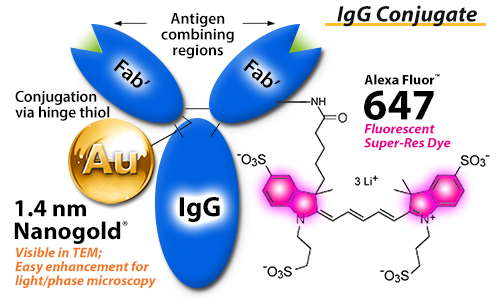
FluoroNanogold™ combined fluorescent and gold nanoparticle immunoprobe
New Fluorescent Label Illuminates Cancer Samples Liu and her team have formulated a new label called Hoechst-Cy5 by combining the DNA-binding molecule Cy5 and a fluorescent dye called Hoechst with ideal blinking properties for superresolution microscopy. Published this week in Science Advances, their study found that this new DNA-binding dye performed well in processed clinical tissue samples ...

Breaking through the Resolution Limit Using Fluorescent Microscopy | Labcompare.com
1.2 Fluorescence Light Microscopy - Cell Structure The addition of fluorescence to light microscopy allows us to look not just at cells, but for things inside them. Specific cellular components can be fluorescently labeled, with a stain or antibody that binds a particular molecule. Alternatively, a protein of interest can be genetically linked to a fluorescent protein such as Green Fluorescent Protein (GFP, isolated from a bioluminescent ...

Label-free fluorescence microscopy offers early cancer detection – Physics World
Fluorescent Labeling - What You Should Know - PromoCell Fluorescent labels offer many advantages, as they are highly sensitive even at low concentrations, are stable over long periods of time, and do not interfere with the function of the target molecules. ... Fluorescence microscopy separates emitted light from excitation light using optical filters. The use of two indicators also allows the ...

FISH - Semrock
Learn about fluorescent labels and light microscopy | Echemi Provide different insights into fluorescent labels and light microscopy on echemi.com. We offer a huge of fluorescent labels and light microscopy news and articles here. ... Fluorescent Brightener. Plastic Rubber Chemicals. Polymer. Precious Metal Catalysts. Zeolite. Flame Retardants. Petrochemical. Natural Products. Lignans. Xanthones ...

Label-free prediction of three-dimensional fluorescence images from transmitted-light microscopy ...
Label-free prediction of three-dimensional fluorescence images from ... Although fluorescence microscopy can resolve subcellular structure in living cells, it is expensive, is slow, and can damage cells. ... phenotypes can be detected via expressed fluorescent labels ...

Light-Sheet Fluorescence Microscopy - Essential Knowledge Briefings
Label-free prediction of three-dimensional fluorescence images from ... Fluorescence microscopy can resolve subcellular structure in living cells, but is expensive, slow, and toxic. Here, we present a label-free method for predicting 3D fluorescence directly from transmitted light images and demonstrate its use to generate multi-structure, integrated images.

FISH - Semrock
The Light Sheet Microscopy Principle - ZEISS The unique Multiview light sheet fluorescence microscope allows you to record the development of large, living samples and gently image them to deliver exceptionally high information content. It is also fast: Lightsheet 7 is your microscope for optical sections at high speed. Acquire images of your whole sample volume at sub-cellular resolution ...

(PDF) Imaging Flies by Fluorescence Microscopy: Principles, Technologies, and Applications
Fluorescence or label-free imaging? Custom microscopy illumination ... For live cell imaging, label-free microscopy is a popular method which comes with several advantages. Unlike fluorescence microscopy, there are no labels that could potentially interfere with the phenomenon you want to observe. It also requires lower light levels, which is great for reducing photodamage.

A guide to light-sheet fluorescence microscopy for multiscale imaging | Nature Methods
Fluorescent Label - an overview | ScienceDirect Topics Fluorescence microscopy is a very common tool. Usually, fluorescent labels are used to brighten up the object of interest. However, the same strategy is not applicable for graphitic materials, such as graphite, graphene, GO or r-GO as they are strong quenchers of dye molecules [45].Therefore, we developed a reverse strategy, FQM, where graphene-based sheets appear dark against a bright ...

Buchmann Institute for Molecular Life Sciences - BMLS
Label-free prediction of three-dimensional fluorescence images from ... Although fluorescence microscopy can resolve subcellular structure in living cells, it is expensive, is slow, and can damage cells. We present a label-free method for predicting three-dimensional fluorescence directly from transmitted-light images and demonstrate that it can be used to generate multi-structure, integrated images.

MIT News: Researchers Improve Electron Microscopy Using Engineered Protein Labels
In Silico Labeling: Predicting Fluorescent Labels in Unlabeled ... - Cell The z-stacks of transmitted-light microscopy images were acquired with different methods for enhancing contrast in unlabeled images. Several different fluorescent labels were used to generate fluorescence images and were varied between training examples; the checkerboard images indicate fluorescent labels that were not acquired for a given example.
Our Services - Charter Preclinical Services | Veterinary Pathology Consulting
Fluorescence Microscopy vs. Light Microscopy - Medical News This light is in the 400-700 nm range, whereas fluorescence microscopy uses light with much higher intensity. The usefulness of traditional light microscopy is hampered by the fact that it uses ...

(a) Tapping-mode AFM image of purified 10 nm NP-A-633 after three... | Download Scientific Diagram
Fluorescent tag - Wikipedia Fluorescent tag. S. cerevisiae septins revealed with fluorescent microscopy utilizing fluorescent labeling. In molecular biology and biotechnology, a fluorescent tag, also known as a fluorescent label or fluorescent probe, is a molecule that is attached chemically to aid in the detection of a biomolecule such as a protein, antibody, or amino acid.

confocal guide 3

Breaking the diffraction limit without fluorescence labels
Post a Comment for "41 fluorescent labels and light microscopy"