41 cell diagram and labels
Animal Cell Diagram | Animal cells worksheet, Animal cell ... - Pinterest Plasma Membrane. Cell Model. Description Teach your students all about the inner working of an animal cell with the help of this hand-drawn animal cell diagram. This pdf packet contains 6 versions of the diagram, to help you teach and also quiz your students: 1. Labeled Animal Cell Diagram - Color 2. Labeled Animal Cell Diagram - Black & White 3. How to Create Venn Diagram in Excel – Free Template Download Replace the default values with the custom labels you previously designed. Right-click on any data label and choose “Format Data Labels.” Once the task pane pops up, do the following: Go to the Label Options tab. Click “Value From Cells.” Highlight the corresponding cell from column Label (H2 for Coca-Cola, H3 for Pepsi, and H4 for Dr ...
Cell Membrane Diagram Labeled : Functions and Diagram Cell Membrane Diagram Labeled Monday, March 22nd 2021. | Diagram Cell Membrane Diagram. There are no organelles in the prokaryotic cells, i.e., they have no internal membrane systems. While lipids help to give membranes their flexibility, proteins monitor and maintain.

Cell diagram and labels
BYJUS BYJUS Plant Cell Diagram | Science Trends A plant cell diagram, like the one above, shows each part of the plant cell including the chloroplast, cell wall, plasma membrane, nucleus, mitochondria, ribosomes, etc. A plant cell diagram is a great way to learn the different components of the cell for your upcoming exam. Plants are able to do something animals can't: photosynthesize. PDF Human Cell Diagram, Parts, Pictures, Structure and Functions One of the few cells in the human body that lacks almost all organelles are the red blood cells. The main organelles are as follows : cell membrane endoplasmic reticulum Golgi apparatus lysosomes mitochondria nucleus perioxisomes microfilaments and microtubules 2
Cell diagram and labels. IXL | Plant cell diagrams: label parts | 8th grade science Identify functions of plant cell parts. O.2. Identify functions of plant cell parts - Eighth grade 2P5. Plant Cells: Labelled Diagram, Definitions, and Structure The cell wall is made of cellulose and lignin, which are strong and tough compounds. Plant Cells Labelled Plastids and Chloroplasts Plants make their own food through photosynthesis. Plant cells have plastids, which animal cells don't. Plastids are organelles used to make and store needed compounds. Chloroplasts are the most important of plastids. CELL MEMBRANE LABEL Diagram | Quizlet identifies or labels the cell. Receptor protein. receives information. Heads. part of the phospholipid that loves water (hydrophili) - points to the most outside and inside of cell. ... Animal Cell Diagram. 14 terms. becker1018 TEACHER. Other Quizlet sets. Med Ethics ch 11. 21 terms. destinyrose3333. BIOL 103 Human Anatomy - LG 21 (Urinary ... A Labeled Diagram of the Plant Cell and Functions of its Organelles A Labeled Diagram of the Plant Cell and Functions of its Organelles We are aware that all life stems from a single cell, and that the cell is the most basic unit of all living organisms. The cell being the smallest unit of life, is akin to a tiny room which houses several organs. Here, let's study the plant cell in detail...
diagram of plant and animal cells to label - TeachersPayTeachers Google Drive™ folder. This worksheet includes both an extensive animal and plant cell diagram to label. The animal cell includes 17 organelles, and the plant cell includes 20 organelles for students to label and color. There is also a 4 page graphic organizer (chart) that includes a drawing of each of the organelles in alphabetical order. Interactive Bacteria Cell Model - CELLS alive In the space are enzymes and other proteins that help digest and move nutrients into the cell. Cell Wall: Composed of peptidoglycan (polysaccharides + protein), the cell wall maintains the overall shape of a bacterial cell. The three primary shapes in bacteria are coccus (spherical), bacillus (rod-shaped) and spirillum (spiral). Animal Cells: Labelled Diagram, Definitions, and Structure The endoplasmic reticulum (s) are organelles that create a network of membranes that transport substances around the cell. They have phospholipid bilayers. There are two types of ER: the rough ER, and the smooth ER. The rough endoplasmic reticulum is rough because it has ribosomes (which is explained below) attached to it. Structure of Cell: Definition, Types, Diagram, Functions - Embibe Various kinds of cells show special differences, yet they all have some basic structural plan consisting of three essential parts: (i) cell membrane ( plasma membrane ), (ii) cytoplasm and (iii) nucleus. Apart from these three components, cells have some living parts that are called cell organelles.
Human Cell Diagram, Parts, Pictures, Structure and Functions One of the few cells in the human body that lacks almost all organelles are the red blood cells. The main organelles are as follows : cell membrane endoplasmic reticulum Golgi apparatus lysosomes mitochondria nucleus perioxisomes microfilaments and microtubules Diagram of the human cell illustrating the different parts of the cell. Cell Membrane Bacteria shapes, structure and diagram - Jotscroll Bacterial spores. Bacterial endospores layers. Bacteria cells are the smallest living cells that are known; even though viruses are smaller than bacteria, viruses are not living cells. There are different types of bacteria with various sizes, shapes, and structures. The bacteria shapes, structure, and labeled diagrams are discussed below. Cell Diagrams - The Biology Corner Open Google Draw and import the diagram. Then use "insert" to create text boxes where students can fill in the labels. Don't forget when assigning this to students on Google classroom to make a copy for each student. You can leave documents in an uneditable form and students can use an addon like "Kami" to annotate the document. Animal Cell Labeled Diagram Pictures, Images and Stock Photos Animal cell Cells consist of a protoplasm enclosed within a membrane, which contains many biomolecules such as proteins and nucleic acids. animal cell labeled diagram stock pictures, royalty-free photos & images. Animal cell. Toxoplasma Gondii Structure Toxoplasma gondii structure.
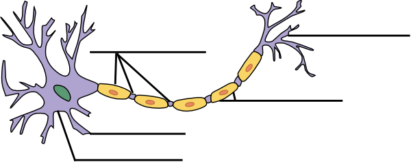
Label The Neuron Clip Art at Clker.com - vector clip art online, royalty free & public domain
Cell Organelles- Definition, Structure, Functions, Diagram Cilia and Flagella are tiny hair-like projections from the cell made of microtubules and covered by the plasma membrane. Structure of Cilia and Flagella Cilia are hair-like projections that have a 9+2 arrangement of microtubules with a radial pattern of 9 outer microtubule doublet that surrounds two singlet microtubules.

ARTimus Prime: 6th Grade- Watercolor Cells
A Labeled Diagram of the Animal Cell and its Organelles A Labeled Diagram of the Animal Cell and its Organelles There are two types of cells - Prokaryotic and Eucaryotic. Eukaryotic cells are larger, more complex, and have evolved more recently than prokaryotes. Where, prokaryotes are just bacteria and archaea, eukaryotes are literally everything else.

Animal Cell Model: What To Use - YouTube
Learn the parts of a cell with diagrams and cell quizzes Labeled cell diagram. For this exercise we'll start with an image of a cell diagram ready labeled. Study this and make sure that you're clear about which structure is found where. Cell diagram unlabeled. It's time to label the cell yourself! As you fill in the cell structure worksheet, remember the functions of each part of the cell that ...
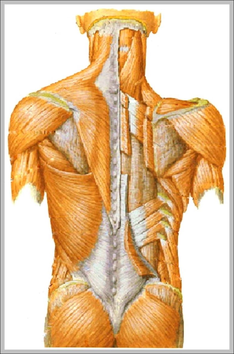
muscular pictures 744×1179 | Anatomy System - Human Body Anatomy diagram and chart images
Images Of Plant Cell Diagram Labeled : Functions and Diagram Images Of Plant Cell Diagram.That's about how the organelles in a cell function. Explore searchView.params.phrase by color family familyColorButtonText(colorFamily.name) plant cell structure - plant cell diagram stock illustrations. comparative illustration of plant and animal cell anatomy (with labels .

Animal Cell by Amy Conley
Skin Diagram with Detailed Illustrations and Clear Labels Skin Diagram The largest organ in the human body is the skin, covering a total area of about 1.8 square meters. The skin is tasked with protecting our body from the external elements as well as microbes.
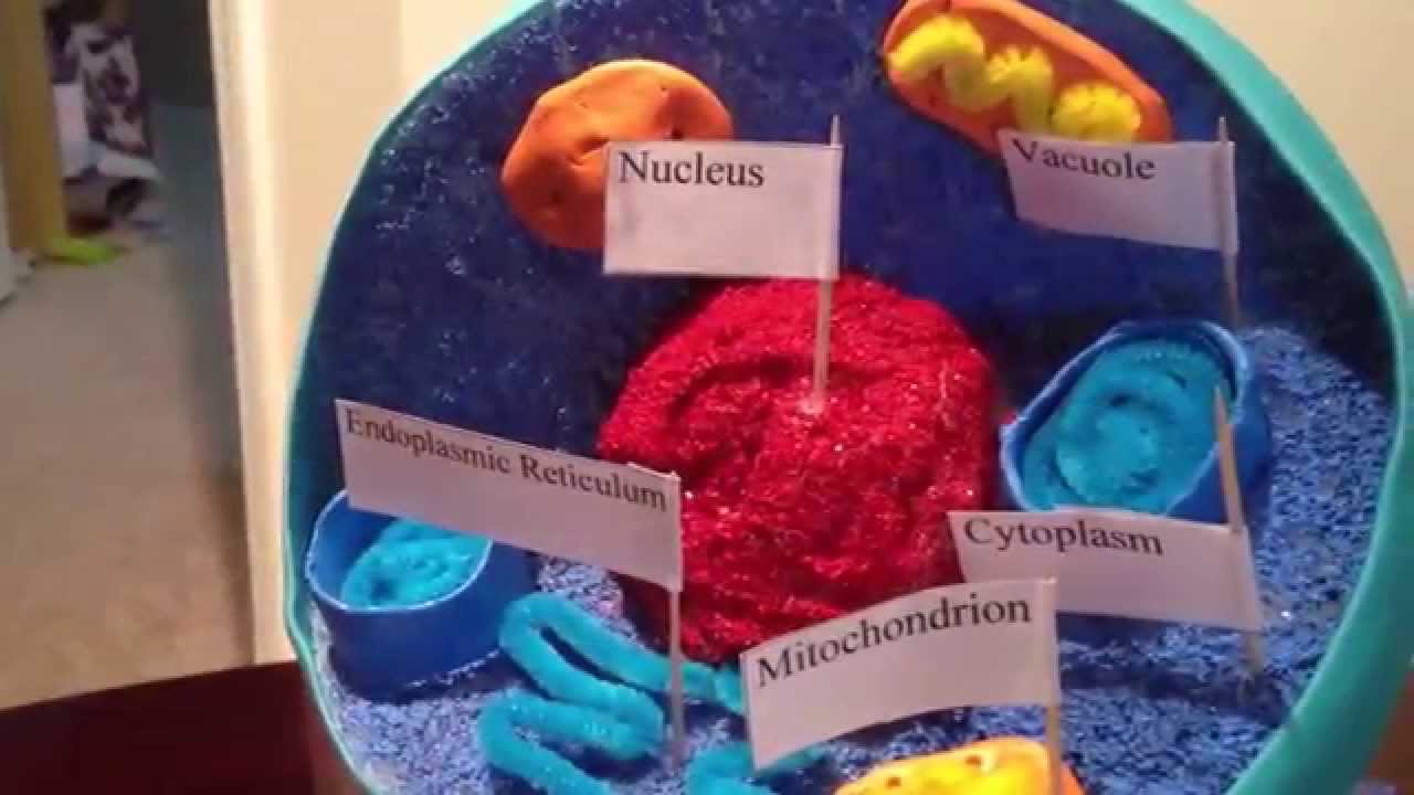
3D Model of Animal Cell - YouTube
How to plot a ternary diagram in Excel Feb 13, 2022 · Adding labels to the apices. Next, we need some space for the apices labels: click into the Plot Area (not the Chart Area) then resize by holding the Shift key (this ensures an equal scaling) and use the mouse cursor on one of the corner pick-points. Then recentre the Plot Area in the Chart Area.
Quia - Protist Vocabulary
Plant and Animal Cell: Labeled Diagram, Structure, Function - Embibe The variation in cell content between plants and animals, as well as their structure and functions, is the source of these differences. Each sensory organ in a cell has a specific job to do. Some cell organelles are found in both plant and animal cells, whereas others are specific to one or the other.

Richard Harwood's Courses: Physical Geography 101: Global Wind Patterns
Label Cell Parts | Plant & Animal Cell Activity | StoryboardThat Student Instructions Create a cell diagram with each part of plant and animal cells labeled. Include descriptions of what each organelle does. Click "Start Assignment". Find diagrams of a plant and an animal cell in the Science tab. Using arrows and Textables, label each part of the cell and describe its function.
Post a Comment for "41 cell diagram and labels"